
Experience advanced 3T MRI imaging for exceptionally clear diagnostic insights today.
3 Tesla MRI
Brain MRI in Wichita, Kansas
A 3 Tesla (3T) MRI represents the most advanced level of magnetic resonance imaging available in
clinical practice, utilizing a stronger magnetic field than standard MRI systems to produce exceptionally detailed, high-resolution images. This increased power allows for greater clarity, improved detection of subtle abnormalities, and enhanced diagnostic confidence.
3T MRI is particularly valuable for imaging the brain, spine, joints, and soft tissues, where precision is critical. It enables physicians to identify conditions earlier and with greater accuracy, often reducing the need for additional testing.
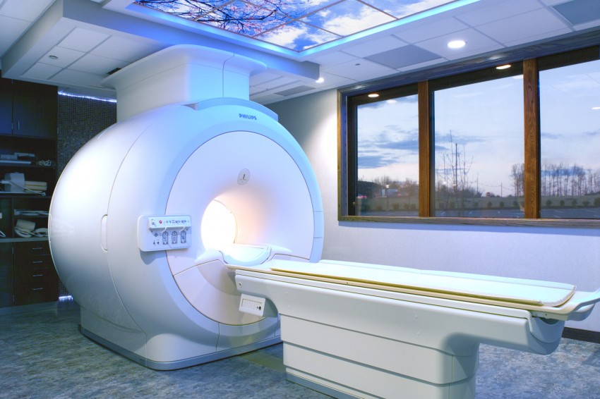
At Elite Diagnostic Imaging, our 3 Tesla MRI technology is designed to deliver superior image quality while maintaining patient comfort. With faster scan times and enhanced detail, combined with interpretation by specialized radiologists, our 3T MRI provides a premium level of diagnostic excellence.
3T MRI: How It Differs From a Standard MRI
Many patients ask, “What is the difference between a 3T MRI and a standard MRI?” The main difference lies in the strength of the magnetic field. A standard MRI typically operates at 1.5 Tesla, while a 3T MRI uses a magnetic field that is twice as strong. This increased strength allows the scanner to capture sharper, more detailed images of the body’s internal structures.
The improved image quality helps physicians detect subtle abnormalities that may be more difficult to identify with lower-strength MRI systems. A 3T MRI is especially beneficial for patients who need a brain MRI in Wichita, Kansas, as well as those requiring detailed imaging of the spine, muscles, ligaments, nerves, and joints, where fine details are essential for an accurate diagnosis. It can also provide faster image acquisition in many cases, helping reduce the amount of time patients spend inside the scanner. While both standard MRI and 3T MRI are safe, non-invasive imaging methods that do not use ionizing radiation, a 3T MRI offers greater diagnostic precision for many complex medical conditions.
3T MRI: What Can You Get?
- Detailed Brain Imaging – Detect neurological conditions such as tumors, stroke, multiple sclerosis, epilepsy, and other brain abnormalities with exceptional image clarity.
- Comprehensive Spine Evaluation – Assess the cervical, thoracic, and lumbar spine for disc injuries, spinal stenosis, nerve compression, and other spinal disorders.
- High-Resolution Joint and Musculoskeletal Imaging – Examine the shoulders, knees, hips, wrists, ankles, and other joints to identify ligament tears, cartilage damage, tendon injuries, and sports-related conditions.
- Soft Tissue and Organ Assessment – Produce highly detailed images of muscles, tendons, ligaments, and selected internal organs to support accurate diagnosis and treatment planning.
- Advanced Vascular and Specialized Imaging – Perform specialized MRI studies, including MR angiography (MRA) and other advanced imaging techniques, to evaluate blood vessels and specific medical concerns without invasive procedures.
Frequently Asked Questions (FAQ)
Ready for Answers? We’re Here to Help.
Take the next step toward clearer answers and greater peace of mind with advanced diagnostic imaging and compassionate care. Contact our team today to schedule your appointment or learn more about the services that best fit your needs.