

Capture detailed internal images quickly for accurate diagnoses and confident treatment decisions.
CT
Computed Tomography in Wichita, Kansas
Computed Tomography (CT) is an advanced imaging technology that uses a combination of X-rays and sophisticated computer processing to create highly detailed, cross-sectional images of the body. At Elite Diagnostic Imaging, we utilize a 64-slice CT scanner, capable of capturing 64 precise images in a single rotation- delivering exceptional speed, clarity, and diagnostic accuracy.
This advanced capability allows for comprehensive visualization of bones, organs, and blood vessels, making computed tomography in Wichita, Kansas, an essential tool for evaluating a wide range of conditions, including injuries, cardiovascular disease, lung disorders, and internal abnormalities. The enhanced detail and rapid imaging also support advanced 3D reconstructions, providing physicians with deeper insight for diagnosis and treatment planning.
Our 64-slice CT technology is designated to maximize image quality while maintaining efficiency and patient comfort.
Every exam is performed by experienced technologists and interpreted by specialized radiologists, ensuring accurate, timely results you and your physician can trust.
CT: How It Works
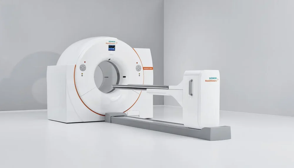
If you’re wondering what happens during a CT scan, the process is generally simple, quick, and comfortable. A CT scanner uses a rotating X-ray source and advanced computer software to capture multiple images from different angles around your body. These images are then combined to create detailed cross-sectional views that allow physicians to examine internal structures without the need for surgery. At Elite Diagnostic Imaging, our specialized radiologists in Wichita, Kansas, carefully interpret these detailed images to help support accurate diagnoses and personalized treatment planning.
During your appointment, you’ll lie on a motorized table that slowly moves through the scanner’s opening. Depending on the type of exam ordered, you may receive a contrast material to improve the visibility of certain organs, blood vessels, or tissues. The scan itself usually takes only a few minutes, although preparation time may vary depending on your specific examination. It is important to remain still during the scan so the images are as clear and accurate as possible.
CT: What Can You Get?
A CT scan can help physicians evaluate many different health concerns by producing highly detailed images of the body’s internal structures. It is commonly used to assess:
- Head, neck, chest, abdomen, and pelvic conditions
- Bone fractures, joint injuries, and spinal abnormalities
- Blood vessels, heart conditions, and circulatory concerns
- Lung diseases, infections, and respiratory conditions
- Tumors, internal bleeding, and other abnormalities requiring detailed imaging
Frequently Asked Questions (FAQ)
Ready for Answers? We’re Here to Help.
Take the next step toward clearer answers and greater peace of mind with advanced diagnostic imaging and compassionate care. Contact our team today to schedule your appointment or learn more about the services that best fit your needs.